A:
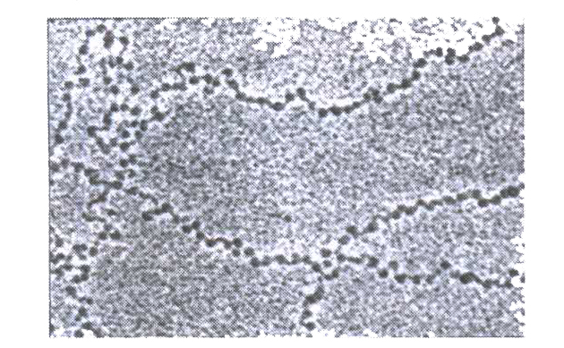
(a) The bead-like structures are the nucleosomes.
1. There is a set of positively charged proteins, called histones, rich in basic amino acid residues, lysine and arginine.
2. Histones are organised to form a unit of eight molecules, called histone octamer.
3. The negatively charged DNA is wrapped around the positively charged histone octamer, to form a nucleosome; a nucleosome contains 200 bp of DNA helix.
(b) The nucleosomes form the repeating units of the chromatin fibre in the nucleus; these nucleosomes of a chromatin are seen as the beads-on-string structure under electron microscope.
1. The chromatin fibres are further coiled and condensed at metaphase stage to form the chromosomes.
2. The packaging of chromatin at higher levels requires another set of proteins, called non-histone chromosomal (NHC) proteins.